This is a 13000 word review published in October 2003, providing a comprehensive overview of the basic and clinical research on hydrogen therapy for various kidney diseases. The writing style of long articles is comprehensive and prone to lacking key points, which is similar to the situation in this article. However, it is still a valuable review. It can provide readers with a comprehensive understanding of all relevant content in a short period of time.
The authors of this review are from Japan's MIZ Corporation, Keio University, and Musashino University School of Data Science.
Hirano S, Ichikawa Y, Sato B, et al. Clinical Use and Treatment Mechanism of Molecular Hydrogen in the Treatment of Various Kidney Diseases including Diabetic Kidney Disease [J] Biomedicines, 2023, 11 (10): 2817
abstract
With the rapid increase of incidence rate of diabetes worldwide, the number of patients with diabetes nephropathy (DKD) has also increased, which is one of the common complications of diabetes. DKD is an important trigger for chronic kidney disease, often leading to end-stage renal failure. However, the effectiveness of medical treatment for DKD still needs to be improved. Hydrogen molecule (H2) is an antioxidant that selectively reduces hydroxyl radicals, making it a highly oxidizing reactive oxygen species. Recent studies have shown that hydrogen not only has antioxidant properties, but also exhibits anti-inflammatory effects, regulating cell lethality and signal transduction. Therefore, it is currently being applied in clinical applications. Many factors contribute to the onset and progression of DKD, among which mitochondrial dysfunction, oxidative stress, and inflammation play important roles. Recent preclinical and clinical trial reports suggest that substances with antioxidant properties may slow down the progression of DKD. Therefore, we conducted a comprehensive review of the literature, focusing on the effectiveness of hydrogen in combating various kidney diseases in animal models and human clinical trials. The evidence collected from this literature review, as well as our previous findings, suggests that hydrogen may bring therapeutic benefits to DKD patients by enhancing mitochondrial function. To confirm these findings, large-scale future clinical studies are needed.
1. Research background
Kidney diseases include many different diseases, such as glomerulonephritis and pyelonephritis caused by kidney inflammation and infection, and kidney disease caused by hypertension, arteriosclerosis and diabetes. Diabetes nephropathy (DKD) is one of the complications of diabetes and the main reason for chronic kidney disease (CKD) to progress to end-stage renal disease (ESRD) [1,2].
The International diabetes Federation estimates that the global prevalence of diabetes will reach 10.5% (53.66 million people) by 2021 and 12.2% (78.32 million people) by 2045 [3]. With the rapid increase of global diabetes, the number of patients with diabetes nephropathy is also rising, especially in developed countries [1,2,3]. Diabetes nephropathy is not only a risk factor for progression to ESRD, but also a risk factor for cardiovascular disease death [4]. About 30-40% of diabetes patients will develop diabetes nephropathy, making it a global public health and health economic problem.
Hydrogen is a flammable, colorless, and odorless gaseous molecule. Hydrogen, as an antioxidant, can directly reduce hydroxyl radicals (· OH) and peroxynitrite (ONOO −), which are reactive oxygen species (ROS) and reactive nitrogen species (RNS), and have very strong oxidation ability [5]. In addition, hydrogen gas also generates a series of indirect effects, including antioxidant, anti-inflammatory, and cytotoxic regulatory effects, by regulating gene expression and through nuclear factor erythroid related factor 2 (Nrf-2) and intracellular signaling.
At present, the total number of research publications on the biological effects of hydrogen has exceeded 2000. Hydrogen not only easily crosses the blood-brain barrier, but also reaches mitochondria through the biofilm, protecting cells from reactive oxygen species - and RNS - induced cell damage. Although the target molecules for hydrogen are not yet clear, recent studies have shown that porphyrin oxide can catalyze the reaction between hydrogen and · OH, thereby alleviating oxidative stress.
The development and progression of diabetes nephropathy involves many factors, among which mitochondrial dysfunction, oxidative stress, hyperglycemia and inflammation have been strongly associated. Recent preclinical and clinical studies have shown that new drugs to reduce oxidative stress may slow down the progress of diabetes nephropathy. Other studies have shown that hydrogen is effective in various animal kidney disease models. In addition, hydrogen inhalation therapy or the use of dialysate rich in hydrogen has been shown to alleviate oxidative stress in dialysis patients. However, as far as we know, the potential therapeutic effect of hydrogen on diabetes nephropathy has not been reported. Therefore, in view of the importance of developing new substances with superior efficacy and safety and showing therapeutic potential for diabetes nephropathy, we hypothesized that hydrogen might alleviate the development and progress of diabetes nephropathy by improving mitochondrial function. Based on this assumption, we aim to analyze the literature on the efficacy of hydrogen gas in various animal kidney disease models and human dialysis patients. In addition, by combining this literature analysis with our previous research on the mechanism of hydrogen against chronic inflammatory diseases [48,49], we explored the potential therapeutic benefits of hydrogen on diabetes nephropathy. We hope that the publication of this article will inspire people to consider the clinical research of hydrogen in the treatment of diabetes nephropathy.
2. Research methods
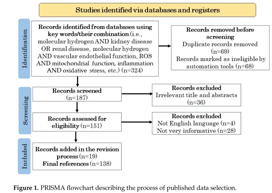
In order to investigate the potential of hydrogen in the treatment of DKD, we conducted a comprehensive literature search, focusing on the etiology and current treatment methods and their efficacy of DKD, the relationship between oxidative stress and inflammation, the production and elimination of reactive oxygen species, the relationship between reactive oxygen species and pathogenesis, mitochondrial function, the role of reactive oxygen species in kidney disease, the impact of hydrogen on animal models and human patients, and the impact of hydrogen on vascular endothelial function. From April 1991 to September 2023, we conducted research using electronic databases from PubMed and Google Scholar. In our PubMed search, we used the Medical Subject Word (MeSH) term. Our search strategy includes using Boolean operators AND, OR, and NOT to combine keywords. The PRISMA flowchart in Figure 1 describes the published method of information selection, following the guidance provided by Page et al. [50,51].
3. Hydrogen regulates oxidative stress
3.1 Research progress of hydrogen in medical applications
In 1975, Dole et al. reported that hydrogen has anti-tumor effects. They found that inhaling a mixture of 2.5% oxygen and 97.5% hydrogen at 8 atmospheres significantly reduced squamous cell carcinoma in mice [52]. In 1994, Abraini et al. demonstrated that inhaling a mixture of 49% hydrogen, 50% helium, and 1% oxygen effectively prevented decompression sickness in divers [53]. In 2001, Ghalib et al. demonstrated the anti-inflammatory effect of high-pressure hydrogen gas on a mouse model of chronic hepatitis caused by schistosomiasis infection [54]. In addition, in 2005, Yanagihara et al. found that saturated neutral hydrogen water produced through water electrolysis reduced chemical oxidant induced liver injury in rats [55].
In 2007, Ohsawa et al. reported the application of hydrogen as a therapeutic antioxidant, which can selectively reduce · OH and ONOO −, two substances with very strong oxidation ability [5]. Since then, the application of hydrogen in medicine has attracted global attention and made significant progress in this field. In 2023, it has been proven that iron porphyrin can catalyze the reaction between hydrogen and · OH [11]. The author proposes a mechanism that, in the presence of hydrogen and porphyrin, the strong oxidative capacity of · OH decreases, activating Nrf-2 as a stimulus like effect and inducing the antioxidant enzyme heme oxygenase-1 (HO-1). However, given that only data on the reaction of iron porphyrins with mammalian tissues or biomolecules appears in the literature, it is clear that the target molecule research for hydrogen is still in its early stages.
3.2 The production and clearance system of reactive oxygen species
Oxygen is necessary for respiratory organisms to produce energy and is used by the mitochondria of cells to produce adenosine triphosphate (ATP). However, 1-2% of the oxygen consumed by the body is converted into reactive oxygen species, which have strong oxidative properties. The main reactive oxygen species in the human body are superoxide anions, hydrogen peroxide gas, · OH, and singlet oxygen. When electrons leak from the mitochondrial respiratory chain and bind with oxygen, superoxide anions are produced. Superoxide anions can also be produced by xanthine oxidase, which uses oxygen and xanthine as substrates, or by a cascade reaction of arachidonic acid in endothelial cells. Superoxide anion is a relatively strong reactive oxygen species, but it is degraded into hydrogen peroxide by superoxide dismutase (SOD). Hydrogen peroxide gas is further decomposed into water and oxygen by catalase (CAT) and glutathione peroxidase (GPX). On the other hand, when oxygen reacts with pigments that act as sensitizers for ultraviolet radiation in the body, singlet oxygen is produced (Figure 2).
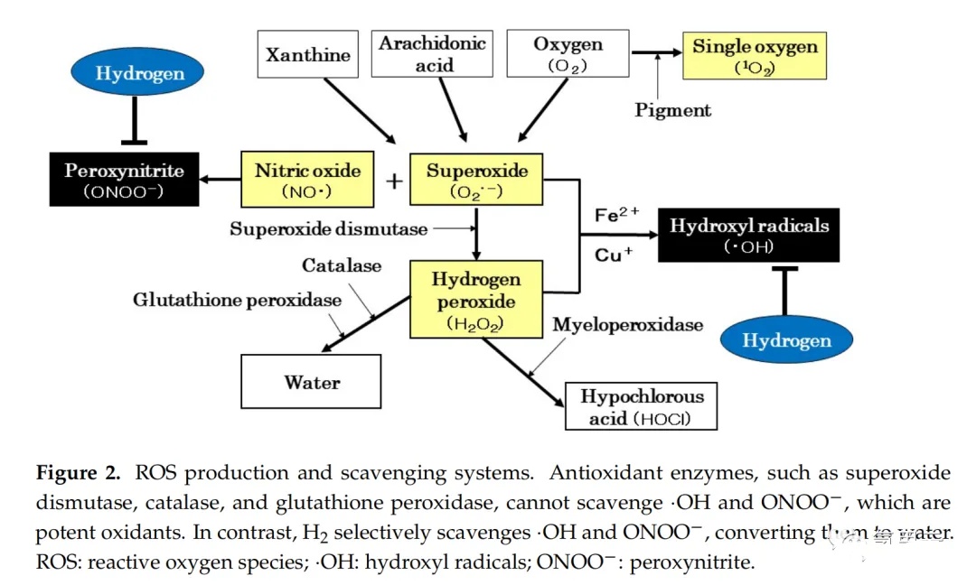
Reactive oxygen species play an important role in the human body, having both physiological functions and contributing to oxidative stress. As age increases, the function of antioxidant enzymes and the body's resistance to reactive oxygen species will gradually weaken. When mental and physiological stress, excessive exercise, smoking, and exposure to ultraviolet and radiation lead to the production of a large amount of reactive oxygen species, the balance between reactive oxygen species and the clearance system is disrupted, and reactive oxygen species that exceed the protective capacity of antioxidant enzymes appear. The imbalance between oxidation and antioxidation can lead to the production of superoxide anions and hydrogen peroxide catalyzed by iron and copper ions, producing · OH, which is a very strong oxidant· OH also exists in other biological reactions and is generated when water, a biological substance, is exposed to radiation· OH only exists in the human body for one millionth of a second, and during this brief period, its oxidation ability is 100 times stronger than that of superoxide anions. On the other hand, nitric oxide (NO ·) reacts with superoxide anions to form ONOO −, which is also a substance with extremely strong oxidative activity. When two strong oxidizing agents, · OH and ONOO −, are produced, they react with nucleic acids, lipids, and proteins in biofilms and tissues, leading to oxidative damage and even DNA oxidation. Antioxidants cannot clear · OH and ONOO −, while hydrogen can selectively reduce · OH and ONOO −, converting them into water (Figure 2).
3.3 The "beneficial" and "harmful" effects of reactive oxygen species
Reactive oxygen species have both harmful and beneficial effects on living organisms. High concentrations of superoxide anions and hydrogen peroxide gas have cytotoxicity, but at low concentrations they serve as second messengers for signal transduction mechanisms and regulatory factors for immune cell metabolism, activation, proliferation, differentiation, and apoptosis. High concentrations of hydrogen peroxide gas can be converted into hypochlorous acid through antioxidant enzymes, thereby protecting the body from bacterial attacks. NO · is crucial for intracellular signal transduction and vascular dilation, and is used clinically as a medical gas. There are reports that oxidative stress can be classified as benign and malignant. A large amount of reactive oxygen species can cause oxidative damage, while a small amount of reactive oxygen species can activate Nrf-2 and induce HO-1, thereby exerting antioxidant effects. A small amount of reactive oxygen species can also induce the expression of tumor suppressor gene p53, which is crucial for preventing cancer. In addition, a small amount of reactive oxygen species is crucial for maintaining the homeostasis of the organism. Oxidative stress caused by reactive oxygen species can lead to mutations in normal cells and promote their transformation into cancer cells. Therefore, the method of removing reactive oxygen species and reducing oxidative stress through antioxidants is considered an effective way to prevent and treat cancer. A large-scale clinical trial of vitamin E supplementation was conducted. However, contrary to expectations, the incidence rate of prostate cancer in patients receiving vitamin E treatment increased significantly. The study conducted using a cancer mouse model also reported the cancer promoting effects of N-acetylcysteine and vitamin E supplementation. DeNicola et al. and Schafer et al. elucidated the mechanism by which these antioxidants promote cancer growth. Therefore, the impact of antioxidants on cancer is two-sided, depending on the conditions. Hydrogen has not yet shown carcinogenic effects, but its carcinogenic inhibitory effect has been confirmed [69].
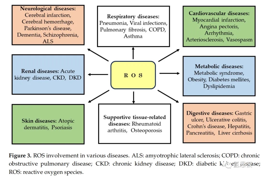
Reactive oxygen species not only play an important role in the occurrence and development of DKD, but also play a crucial role in many other diseases. These include neurological disorders such as cerebral infarction, cerebral hemorrhage, Parkinson's disease, dementia, schizophrenia, and amyotrophic lateral sclerosis; Cardiovascular diseases such as myocardial infarction, angina, arrhythmia, arteriosclerosis, and vasospasm; Respiratory system diseases such as pneumonia, viral infections, pulmonary fibrosis, chronic obstructive pulmonary disease, and asthma; Kidney diseases such as acute kidney injury, CKD, and DKD; Digestive system diseases such as gastric ulcer, ulcerative colitis, Crohn's disease, hepatitis, pancreatitis, and cirrhosis; Metabolic diseases such as metabolic syndrome, obesity, diabetes and dyslipidemia; Skin diseases such as eczema and psoriasis; And support for tissue related diseases such as rheumatoid arthritis and osteoporosis. Therefore, it is not an exaggeration to say that reactive oxygen species are involved in most diseases that affect various organs and tissues of the human body (Figure 3).
The role of mitochondria in renal diseases
4.1 The structure and function of mitochondria
Mitochondria are organelles that produce over 90% of cellular energy and generate ATP through oxidative phosphorylation (OXPHOS) under aerobic conditions. Mitochondria are composed of the outer membrane, the inner membrane, the gaps between the inner and outer membranes, the matrix surrounded by the inner membrane, and the cristae surrounded by the sunken inner membrane. Mitochondria have their own genome, namely mitochondrial DNA (mtDNA), which is different from the nuclear genome (nDNA).
About 2 billion years ago, aerobic bacteria Proteobacteria invaded archaea, transforming them into mitochondria. Therefore, the structure of mtDNA is similar to that of the bacterial genome, rather than the nuclear genome of eukaryotic cells. The mitochondrial respiratory chain complex I-V is assembled in the inner membrane crest to efficiently generate ATP. During this process, a large amount of mitochondrial reactive oxygen species (mtROS) are mainly generated from complexes I and III, which are usually cleared by the antioxidant system. Therefore, mitochondria maintain efficient energy production through the electron transfer system of the inner membrane respiratory chain complex and the OXPHOS of ATP synthase.
4.2 The role of reactive oxygen species in renal diseases
Mitochondrial diseases are caused by various genetic abnormalities, which are involved in the maintenance of mitochondrial function and structure, including ATP synthesis, amino acid, lipid and protein transport, and clearance of oxidative stress within mitochondria. These genetic abnormalities can be attributed to abnormalities in mtDNA and genetic mutations in nDNA [76]. When mitochondrial function is impaired due to these genetic abnormalities, organs with high energy demands will experience functional impairment and exhibit various symptoms. Mitochondrial diseases are called mitochondrial encephalomyopathy or myopathy based on the location of the lesion. In addition to genetic abnormalities that lead to reduced ATP production and increased mitochondrial reactive oxygen species, there are other factors that can induce abnormal mitochondrial function, and when damaged mitochondria release apoptotic signals such as cytochrome c, it can lead to cell death. This type of mitochondrial dysfunction has been observed in many metabolic and neurodegenerative diseases. Among them, glomerular capillary endothelial cells, podocytes that perform glomerular filtration function, and renal tubular epithelial cells are active in mitochondrial metabolism, making the kidneys susceptible to energy metabolism failures caused by ischemia, hypoxia, and toxic substances. With the increase of the number of patients with diabetes, DKD has become the main disease of ESRD [1]. Diabetes is considered to be the main reason for the recent increase in the number of dialysis patients. It reduces the insulin action in cells, leading to chronic hyperglycemia and abnormal blood lipids. Therefore, abnormalities in signal transduction mechanisms can be observed, such as nutrient sensitive AMP activated protein kinase (AMPK) and rapamycin target protein complex 1, as well as mitochondrial dysfunction. In addition, due to excessive intracellular glucose intake, diabetes also increases oxidative stress, which promotes mitochondria to produce excessive mt reactive oxygen species, activate the polyol metabolic pathway, activate protein kinase C, and lead to the accumulation of advanced glycation end products. Oxidative stress also promotes the development of DKD. However, physiological levels of reactive oxygen species are crucial for regulating intracellular signaling mechanisms and cellular homeostasis. In diabetes, mitochondrial membrane potential and respiratory regulation rate decrease, and vice versa, the production of reactive oxygen species decreases, which inhibits the production of OXPHOS and ATP. The sustained decrease in OXPHOS and ATP production in mitochondria increases oxidative stress outside the mitochondria due to the uncoupling of nicotinamide adenine dinucleotide phosphate (NADPH) oxidase and endothelial NO · synthase, releasing cytokines involved in inflammation and fibrosis and inducing cell damage. In this case, reactive oxygen species from the cytoplasm play a more important role than mt reactive oxygen species. Previous studies on the use of AMPK activators in diabetes animal models have demonstrated that they can improve mitochondrial function and induce the production of mt reactive oxygen species, thereby reducing proteinuria and reducing DKD. As part of the mitochondrial targeted therapy strategy for kidney disease, a therapeutic approach has been proposed to improve mitochondrial function and induce physiological levels of reactive oxygen species production, which has practical value.
4.3 Development of drugs for diabetes nephropathy
As a potential therapeutic drug for diabetes nephropathy (DKD), badosolone methyl (BM) has attracted the most attention. Badosolone methyl is a novel synthetic triterpenoid compound with an oleic acid backbone. Badosolone methyl was initially developed as an anti-cancer drug. In the first phase clinical trial of cancer patients, it was found that the estimated glomerular filtration rate (eGFR) increased, therefore, badosolone methyl was converted into a DKD treatment drug [94]. The main mechanism by which Badosolon methyl improves renal function is by activating the Kelch like ECH related protein 1 (Keap1)/Nrf-2 pathway. After conducting a phase II open label and double-blind placebo-controlled trial in Japan, a large-scale double-blind placebo-controlled trial (AYAME study) was conducted to demonstrate the efficacy and safety of batosolon methyl. However, although this clinical trial showed improvement in eGFR, it failed to demonstrate that batosolone methyl inhibited the development of ESRD, thus further research on batosolone methyl was terminated. According to reports, other substances with a mechanism similar to that of batosolone methyl include curcumin, isothiocyanates, cinnamaldehyde, resveratrol, and alpha lipoic acid. These substances are in the preclinical or early clinical trial stage and are expected to serve as activators of the Keap1/Nrf-2 pathway in the future. In addition, sodium glucose transporter 2 (SGLT2) inhibitors act through oxidative stress and inflammation, while glucagon like peptide-1 (GLP-1) receptor agonists act on the AMPK mammalian target mTOR autophagy reactive oxygen species signaling axis, both of which have been shown to be effective in animal models of DKD. These SGLT2 inhibitors and GLP-1 receptor agonists are expected to become therapeutic drugs against oxidative stress. On the other hand, DKD therapeutic drugs have been developed to improve mitochondrial function. A highly targeted antioxidant for mitochondria is coenzyme Q (MitoQ), which has been found to be effective in animal models of I/R injury and DKD. However, MitoQ has many limitations, such as becoming an oxidant at high doses, thereby reducing its efficacy. Elemiprid (SS-31), a cardiolipin acting on the inner membrane of mitochondria, is also effective in I/R injury and DKD models. Multiple clinical trials of SS-31 in DKD patients are currently underway. MA-5 is a newly synthesized indole compound characterized by its ability to increase intracellular ATP and reduce mt reactive oxygen species, which has also attracted people's interest. MA-5 is effective in treating I/R nephropathy and cisplatin induced nephropathy. It also improves the respiratory function of mitochondria in the heart and kidneys, prolongs the lifespan of mice with mitochondrial diseases, and is currently undergoing clinical trials in patients with mitochondrial diseases. In addition, plants and their extracts, as well as vitamins, have shown potential therapeutic potential against DKD due to their ability to regulate oxidation and inflammation, which are key factors in the development and progression of DKD. For example, essential oils from herbs such as Embelia ribes Burm have been reported to have antioxidant properties. Vitamin C and E, allicin in garlic and procyanidins extract in grape seeds also have antioxidant, anti-inflammatory and/or anti diabetes effects. These candidate drugs are in the preclinical research stage; Therefore, we have high expectations for future research and development.
5. The effects of hydrogen on various kidney diseases and vascular endothelial function
Here, we investigated the relevant literature on the preventive and therapeutic effects of hydrogen in animal kidney disease models and human kidney diseases. The following is a summary of studies reporting the effects of hydrogen on I/R injury, transplantation, CKD, drug-induced kidney injury, kidney stones, renal fibrosis, sepsis related acute kidney injury (AKI), peritoneal dialysis (PD), and hemodialysis (HD) (Table 1). Normal endothelial cells can dilate and contract blood vessels, proliferate and inhibit proliferation of vascular smooth muscle cells, coagulation and anticoagulant blood, inflammation and anti-inflammatory, oxidative and antioxidant properties. The balance of these opposing effects maintains vascular tension and regulates and maintains vascular structure. However, oxidative stress from reactive oxygen species and RNS can induce vascular endothelial damage, which is one of the factors in the development of atherosclerosis. In addition, this atherosclerosis is a risk factor for CKD (including DKD) and cardiovascular disease. Therefore, this chapter also provides a literature review on the effects of hydrogen on vascular endothelial function.
5.1 Impact on animal kidney disease models
5.1.1 Ischemia reperfusion injury
Renal I/R injury is an important cause of AKI and one of the factors contributing to the development of CKD. Shingu et al. investigated the protective effect of hydrogen rich saline solution (HRS) on renal I/R injury in rats. Hydrogen saline improved mitochondrial morphology and significantly reduced blood urea nitrogen (BUN), creatinine (Cr), and 8-hydroxydeoxyguanosine (8-OHdG). Similarly, in a rat kidney I/R injury model, Wang et al. found that hydrogen saline significantly inhibited BUN, Cr, malondialdehyde (MDA), 8-OHdG, tumor necrosis factor alpha (TNF - α), interleukin-1 β, IL-6, and myeloperoxidase (MPO), and significantly increased tissue superoxide dismutase (SOD) and catalase (CAT) activity. Li et al. also studied the effect of hydrogen saline on a rat renal I/R injury model, and the results showed that hydrogen saline significantly reduced stromal congestion, edema, and bleeding in renal tissue, as well as BUN, Cr, B-cell lymphoma 2 (Bcl-2), caspase-3, -8 and -9, IL-6, and TNF - α, while Bcl-2 associated x protein (Bax) significantly increased [23]. In addition, they reported that the protective effect of hydrogen saline may be attributed to its anti apoptotic and anti-inflammatory effects. Chen et al. investigated the protective effect of hydrogen saline in an I/R induced AKI mouse model, and the results showed that it significantly reduced renal tissue fibrosis, BUN, and Cr, and increased the level of the anti-aging gene Klotho [24]. In addition, they demonstrated that hydrogen saline increased the damage regulating autophagy regulator (Beclin-1) and microtubule associated protein light chain 3-II (LC3-II). They believe that hydrogen saline exerts its protective effect by maintaining Klotho expression and activating autophagy in the kidneys. Xu et al. investigated the effect of hydrogen saline on a rat kidney I/R injury model and reported that hydrogen saline significantly reduced BUN, Cr, MDA, and 8-OHdG, and increased heme oxygenase-1 (HO-1) gene expression and SOD activity [25]. They believe that hydrogen saline improves renal I/R injury in rats by reducing oxidative stress and increasing HO-1 gene expression.
5.1.2 Kidney transplantation
Reactive oxygen species participate in the development of chronic allogeneic kidney transplantation (CAN), leading to interstitial fibrosis and tubular atrophy. Cardinal et al. investigated the effect of drinking hydrogen rich water on a rat allogeneic kidney transplantation model and found that hydrogen rich water slowed down CAN progression, decreased MDA, TNF - α, and IL-6 by reducing BUN, Cr, and urinary protein, further prolonging overall survival and improving transplanted kidney function [26]. Hydrogen rich water also reduces the activation of inflammatory signaling pathways, such as mitogen activated protein kinase (MAPK), indicating its effectiveness in preventing CAN and prolonging kidney transplant survival. I/R injury is inevitable in kidney transplantation, affecting both short-term and long-term transplant survival rates. Abe et al. investigated the inhibitory effect of hydrogen rich University of Wisconsin (HRUW) solution on I/R injury in a rat kidney allograft model. The results showed that it reduced MDA and 8-OHdG in the kidney allograft, as well as the number of TUNEL stained cells and ED-1 positive cells in the renal tubules [27]. It also reduces Cr and urinary protein, thereby improving renal function and prolonging the survival of receptors, indicating that HRUW reduces tubular injury and thus reduces the development of interstitial fibrosis. On the other hand, AKI has a significant impact on the survival rate of liver transplant recipients. Du et al. investigated the protective effect of hydrogen saline on AKI after orthotopic liver transplantation in rats, and the results showed that hydrogen rich water reduced tissue damage and lowered BUN, Cr, MDA, and SOD [28]. Meanwhile, hydrogen saline significantly improved apoptosis and inhibited the expression of caspase-3 and cytochrome c. In addition, the expression of Beclin-1 and LC3-II was upregulated [28]. Chloroquine, as an autophagy inhibitor, counteracts the protective effect of hydrogen salt water [28]. These findings indicate that hydrogen saline prevents AKI by reducing apoptosis and activating autophagy.
5.1.3 Chronic kidney disease
Dahl salt sensitive (SS) rats are a CKD model animal that develops hypertension and kidney damage with age. Zhu et al. investigated the effect of electrolyzed hydrogen water (EW) on ischemia induced cardiac and renal injury in Dahl SS rats [29]. Rats were fed with EW or filtered water (FW) and then underwent unilateral renal I/R. The control group of rats receiving FW showed a significant increase in BUN, methylglyoxal, and MCP-1 [29]. In the histological examination of the kidneys and hearts, the nitrotyrosine staining of the control group rats significantly increased [29]. However, these findings showed significant improvement in rats treated with EW, indicating that EW has the potential to prevent CKD [29]. Zhu et al. also studied the effects of EW and FW on age-related cardiac and renal injury in Dahl SS rats [30]. Urinary protein and cardiac remodeling increased in the FW group. Histologically, significant age-related changes were observed in the kidneys and heart; However, these changes were significantly reduced in rats treated with EW, with decreased MDA and nitrotyrosine levels [30]. Xin et al. investigated the protective effect of hydrogen rich water on renal injury in spontaneously hypertensive rats (SHRs). Hydrogen rich water significantly reduced the BUN and Cr of SHRs, reduced the production of reactive oxygen species, increased SOD, GPX, and CAT activities, and inhibited NADPH oxidase activity [31]. It also inhibits the expression of TNF - α, IL-6, and IL-1 β. In addition, hydrogen rich water has an improving effect on mitochondrial morphology and function, including inhibiting the production of reactive oxygen species and mitochondrial swelling, and increasing ATP production [31].
5.1.4 Drug induced renal injury
Cisplatin is a widely used anticancer drug for the treatment of various tumors; However, its application is limited by the nephrotoxicity caused by oxidative stress. Nakashima Kamimura et al. reported that when mice inhaled hydrogen or drank hydrogen rich water, hydrogen reduced kidney damage while not affecting the anti-tumor activity of cisplatin [32]. In other words, hydrogen or hydrogen rich water improved cisplatin induced mortality and weight loss, improved kidney tissue damage, and restored Cr and BUN [32]. Li et al. investigated the therapeutic effect of hydrogen rich water in a rat model of iron nitrotriacetic acid induced kidney injury [33]. Hydrogen rich water reduces the production of Cr, BUN, MDA, ONOO − and NADPH oxidase activity, while increasing CAT activity. Hydrogen rich water improves mitochondrial dysfunction and oxidative stress, including renal mitochondrial swelling, reduced ATP production, and increased production of reactive oxygen species [33]. Hydrogen rich water also inhibits inflammation, manifested by decreased expression of NF - κ B, IL-6, and MCP-1 in the kidneys [33]. In addition, hydrogen rich water inhibits the expression of vascular endothelial growth factor (VEGF) and phosphorylation of signal transduction and transcriptional activator 3 (STAT3), thereby reducing the incidence of renal cell carcinoma and inhibiting tumor growth [33]. Oxidative stress caused by cyclosporine A is one of the main causes of chronic kidney injury. Lu et al. studied the alleviating effect of hydrogen rich water on cyclosporine A induced kidney injury in rats and found that it reduced reactive oxygen species production, MDA and Keap1 levels, and increased the expression of Nrf-2 and HO-1 [34]. They propose that the role of hydrogen rich water involves improving oxidative stress by activating the Keap1/Nrf-2 signaling pathway.
5.1.5 Kidney stones
Peng et al. evaluated the protective effect of hydrogen on glyoxylate induced renal calcium oxalate (CaOx) crystallization deposition in mice and reported that it reduced MDA and 8-OHdG levels, increased SOD, GSH, and CAT activity [35]. They also showed that hydrogen reduced MCP-1 levels and increased IL-10 expression, indicating that hydrogen played a protective role by reducing renal crystallization, renal oxidative damage, and inflammation [35].
5.1.6 Renal fibrosis
Xu et al. investigated the therapeutic effect of hydrogen saline in a rat model of unilateral ureteral obstruction (UUO) induced renal fibrosis, and the results showed that it significantly improved renal injury score, cell apoptosis index, matrix fibrosis, and macrophage infiltration in renal tissue [36]. In addition, hydrogen salt water reduced MDA levels and increased SOD activity.
Furthermore, Xing et al. investigated the therapeutic effect of hydrogen rich water in a mouse model of renal fibrosis induced by UUO, and the results showed that it inhibited Cr, BUN, and renal fibrosis [37]. They also studied the inhibitory effect of hydrogen rich water on renal epithelial mesenchymal transition (EMT) induced by transforming growth factor - β 1 (TGF - β 1) using human renal proximal tubular epithelial cells. The results showed that hydrogen rich water eliminated EMT and restored reduced expression of sirtuin-1 (Sirt1) [37]. The inhibitor Sirtinol of Sirt1 eliminated the inhibitory effect of hydrogen rich water on EMT, indicating that hydrogen rich water improves kidney injury and fibrosis by regulating Sirt1 [37].
Congenital obstructive kidney disease is a common pathophysiological cause of CKD, and the release of reactive oxygen species can exacerbate renal fibrosis. Shuiye et al. evaluated the therapeutic effect of hydrogen rich water in a rat UUO induced kidney injury model [38]. Hydrogen rich water inhibits tubulointerstitial injury, reduces the area of interstitial fibrosis, and the frequency of TGF - β 1 positive cells [38]. In addition, hydrogen rich water restored the decrease in Klotho mRNA expression.
5.1.7 Sepsis induced renal injury
Liu et al. investigated the combined effect of early infusion resuscitation and hydrogen gas on AKI induced by lipopolysaccharide in septic shock rats, and the results showed that the combination of the two reduced BUN and Cr [39]. It also reduces MDA and reduces renal levels of TNF - α and IL-6 compared to intravenous resuscitation alone [39]. These findings indicate that early infusion resuscitation combined with hydrogen gas has a stronger protective effect on AKI.
Yao et al. examined the protective effect of inhaling hydrogen in a sepsis related AKI model induced by cecal ligation and puncture in mice [40]. AKI occurs in the early stages of sepsis, characterized by an increase in BUN and Cr, renal fibrosis, increased apoptosis of renal tubular epithelial cells, as well as macrophage infiltration and production of inflammatory cytokines (IL-6 and TNF - α) [40]. In contrast, hydrogen salt inhalation increased the mRNA levels of anti-inflammatory cytokines (IL-4 and IL-13) in renal tissue and enhanced the production of anti-inflammatory cytokines (IL-10 and TGF - β), indicating that hydrogen inhalation is effective in protecting the kidneys and reducing inflammation in septic AKI [40].
5.1.8 other
Guo et al. investigated the efficacy of hydrogen water in an early AKI model induced by severe burns in rats, and reported that hydrogen rich water improved renal function (BUN and Cr) and reduced tubular apoptosis [41]. In addition, the mechanism by which hydrogen rich water improves AKI involves inhibiting oxidative stress-induced apoptosis and inflammation, which seems to be achieved by regulating the MAPK and nuclear factor (NF) - κ B signaling pathways [41].
Shi et al. investigated the protective effect and potential mechanism of hydrogen rich water on AKI in acute pancreatitis induced by taurocholic acid in rats [42]. They found that hydrogen rich water prevented the progression of inflammatory cascade reactions and alleviated oxidative damage to the kidneys by inhibiting NF - κ B activation and clearing reactive oxygen species [42].
In addition, Guan et al. analyzed the protective effect of hydrogen on renal injury caused by chronic intermittent hypoxia (CIH) in rats, including oxidative stress, autophagy, and endoplasmic reticulum stress [43]. Hydrogen also improved the renal function of CIH rats, reducing tissue damage, oxidative stress, and apoptosis. They found that hydrogen alleviated CIH induced kidney injury by inhibiting oxidative stress-dependent MAPK activation, thereby reducing endoplasmic reticulum stress and activating autophagy [43].
5.2 The impact of hydrogen on human kidney disease
5.2.1 peritoneal dialysis
Oxidative stress caused by glucose degradation products is the cause of peritoneal degradation in peritoneal dialysis patients. Terawaki et al. investigated the effect of hydrogen rich dialysate (HED) on peritoneal oxidative stress in six peritoneal dialysis patients [44]. According to the results, compared to patients receiving standard dialysis fluid treatment, patients receiving HED treatment had a higher percentage of reduced albumin in their dialysis fluid and serum, and a lower percentage of oxidized albumin. Therefore, HED seems to alleviate oxidative stress in the peritoneum and whole body [44].
5.2.2 Hemodialysis
Nakayama et al. developed a dialysis system using dialysate dissolved in hydrogen gas and studied the efficacy of HED in 21 hemodialysis patients. HED significantly reduced systolic blood pressure before and after dialysis [45]. In addition, it significantly reduced MCP-1 and MPO, indicating its potential to control uremia by reducing inflammation [45]. Terawaki et al. conducted a cross study using standard dialysate (SD) and HED to investigate the effect of HED on oxidative stress in eight hemodialysis patients. The results showed that HED significantly reduced the average percentage of oxidized albumin in serum at the outlet of the dialysis system, higher than SD [46]. Sokawa et al. investigated the effects of hydrogen on oxidative stress and inflammatory response in six hemodialysis patients [47]. Inhaling hydrogen gas three times a week for two weeks did not affect the biological antioxidant potential (BAP). However, it significantly reduced dichlorofluorescein reactive oxygen species (d-ROMs) and C-reactive protein (CRP), and these effects persisted for two weeks after discontinuing hydrogen inhalation, indicating that hydrogen inhalation can alleviate oxidative stress and inflammatory response in hemodialysis patients [47].
5.3 The impact on endothelial function of blood vessels
Jiang et al. induced damage to cultured rat vascular endothelial cells by adding advanced glycation end products (AGEs) and examined the protective effect of hydrogen medium (HRM) [113]. They showed that HRM significantly reduces reactive oxygen species, increases antioxidant enzymes, and reduces apoptosis [113]. These results indicate that hydrogen inhibits AGE induced endothelial injury by inhibiting oxidative stress and apoptosis. Ohsawa et al. studied the inhibitory effect of hydrogen rich water on atherosclerosis by letting apolipoprotein E (ApoE) deficient mice drink hydrogen rich water [114]. They showed that the atherosclerotic lesion area of mice in the hydrogen rich water group was significantly reduced [114]. In addition, it was observed that the accumulation of macrophages in the arterial tissue of mice was inhibited and the level of oxidative stress was reduced, which indicates that drinking hydrogen rich water may prevent atherosclerosis in ApoE deficient mice [114]. Truong et al. investigated the effect of hydrogen on endothelial glycocalyx in a heat stroke induced rat model [115]. They showed that hydrogen increased the survival rate of heat stroke induced rats and inhibited the shedding of endothelial glycocalyx. In addition, hydrogen reduces MDA and TNF - α levels, while increasing SOD levels [115]. These results indicate that hydrogen reduces endothelial glycocalyx damage through antioxidant and anti-inflammatory effects.
Song et al. investigated the effect of hydrogen rich water on improving lipid metabolism in 20 patients with metabolic syndrome [116]. They conducted a comparative experiment using high-density lipoprotein (HDL) in serum collected from patients before and after drinking hydrogen rich water. They demonstrated that drinking hydrogen rich water inhibited the oxidation of low-density lipoprotein (LDL), inhibited monocyte adhesion to endothelial cells, promoted cholesterol extraction from macrophages that uptake oxidized LDL, and inhibited endothelial cell apoptosis [116]. These results indicate that hydrogen rich water improves lipid metabolism by improving HDL function in endothelial cells.
Sakai et al. investigated the effect of hydrogen rich water on endothelial function in healthy subjects [117]. They evaluated endothelial function by measuring blood flow mediated dilation (FMD) of arteries. Comparing the FMD before and after taking placebo water or hydrogen rich water alone, it was found that 18 subjects in the placebo group had a decrease in FMD, while 16 subjects in the hydrogen rich water group had an increase in FMD. This change is a significant improvement in endothelial function [117]. These results indicate that hydrogen may maintain NO mediated vasodilation and contraction reactions.
Ishibashi et al. also investigated the effect of continuous consumption of hydrogen rich water for two weeks on endothelial function in healthy subjects in a randomized controlled trial [118]. They used peripheral arterial tension measurement to measure the reactive congestion index (RHI) of finger blood vessels to evaluate endothelial function. The results showed that the RHI of the hydrogen rich water group (34 subjects) was significantly higher than that of the placebo group (34 subjects), especially 24 hours after the first consumption of hydrogen rich water and two weeks after continuous consumption of hydrogen rich water [118]. These results indicate that hydrogen improves the endothelial function of arteries or small arteries.
In general, this literature review shows that hydrogen may prevent endothelial dysfunction and prevent diabetes nephropathy from developing into cardiovascular disease by reducing oxidative stress, inflammation and apoptosis.
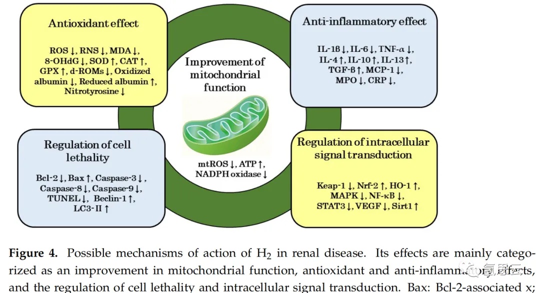
Many studies have shown that hydrogen has a good preventive and therapeutic effect on kidney diseases. It can improve tissue damage, reduce serum BUN and Cr, and urinary protein. According to the literature review, the mechanism of action of hydrogen in kidney disease can mainly be divided into improving mitochondrial function, antioxidant and anti-inflammatory effects, as well as regulating cell death and intracellular signal transduction. These mechanisms are not independent of each other, but rather complex mechanisms that interact to form hydrogen gas (Figure 4).
6.1 Improving mitochondrial function
Transmission electron microscopy is a commonly used method to examine the morphological changes of mitochondria. Research on the effect of hydrogen on mitochondrial morphology has shown that hydrogen can alleviate mitochondrial swelling [21,31]. It can also inhibit the production of reactive oxygen species in mitochondria, enhance ATP production, and reduce NADPH oxidase activity [31,33], indicating that its mechanism of action involves the improvement of mitochondrial morphology and function.
6.2 Antioxidant effects
Using fluorescent reagents, hydrogen has been shown to reduce the fluorescence intensity of reactive oxygen species or RNS in renal tissue. Hydrogen can also reduce the markers of lipid peroxidation, MDA, and DNA oxidation, 8-OHdG. The activity of antioxidant enzymes such as SOD, CAT, and GPX in renal tissue is used as markers of antioxidant activity; Hydrogen increases the activity of these antioxidant enzymes. In addition, oxidative proteins and d-ROMs are used as oxidative markers in human clinical trials, while reducing proteins and BAP are used as antioxidant markers; Hydrogen gas reduces oxidative proteins and d-ROMs, and increases the content of reducing proteins in PD or HD patients. On the other hand, ONOO − can modify the tyrosine residues exposed on the protein surface to produce nitrotyrosine. This nitrotyrosine has attracted attention as it is an oxidative stress marker in various inflammatory diseases; Hydrogen reduces ONOO − and inhibits the production of nitrotyrosine, thereby improving oxidative stress, nitro stress, and inflammation [29,30]. Therefore, the experimental results indicate that hydrogen improves kidney damage through its antioxidant properties.
6.3 Anti inflammatory effects
IL-1 β, IL-6, and TNF - α are inflammatory factors, while IL-4, IL-10, IL-13, and TGF - β are anti-inflammatory factors. In experiments examining the mRNA expression or protein levels of these factors in these animal models, hydrogen reduced the levels of the former while increasing the levels of the latter. In addition, MCP-1 and MPO have been used as markers for macrophage infiltration and inflammatory response. In animal models of kidney disease and HD patients, hydrogen reduced the levels of MCP-1 and MPO, and in human HD patients, CRP levels were reduced [47]. These findings indicate that hydrogen has a protective effect on kidney damage through its anti-inflammatory properties.
6.4 Regulation of cell lethality
Bcl-2 is a protein that promotes cell apoptosis, while Bax is a protein that inhibits cell apoptosis. The protease family involved in cell apoptosis includes caspase-3, -8, and -9. In addition, the TUNEL staining method is used to detect DNA fragmentation caused by cell apoptosis. In a kidney disease model, hydrogen not only inhibited the expression of Bcl-2 gene and increased the expression of Bax gene, but also inhibited the expression of caspase-3, -8, and -9 [23,28]. Hydrogen also reduces TUNEL positive cells in renal tubules [27]. On the other hand, Beclin-1 and LC3-II have been identified as regulators and markers of autophagy; Hydrogen not only improves kidney damage, but also increases the expression of Beclin-1 and LC3-II [24,28]. However, the autophagy inhibitor chloroquine counteracted the effect of hydrogen gas [28]. These findings indicate that hydrogen regulates cell lethality by inhibiting cell apoptosis and activating autophagy.
6.5 The regulatory role of signal transduction
Previous studies have found that the protective mechanism of hydrogen on kidney disease involves a decrease in Keap1 levels and an increase in Nrf-2 and HO-1 gene expression [25,34]. Therefore, the activation of the Keap1/Nrf-2 signaling pathway to alleviate oxidative stress and enhance biological defense function is involved in the action of hydrogen. In addition, studies have shown that inhibiting signaling pathways such as MAPK and NF - κ B plays a role in the antioxidant and anti-inflammatory effects of hydrogen gas. In addition, the signaling pathways that inhibit STAT3 phosphorylation and VEGF expression, as well as the signaling pathways that activate Sirt1 expression, also contribute to the effect of hydrogen. Due to the counteraction of hydrogen by Sirtinol (a Sirt1 inhibitor), Sirt1 may be involved in the protective effect of hydrogen on kidney disease [37]. Therefore, hydrogen exerts its improving effect on kidney injury by activating or inhibiting various signaling pathways.
7. The potential of hydrogen in the treatment of diabetes nephropathy
7.1 Therapeutic potential of hydrogen in the pathogenesis of diabetes nephropathy
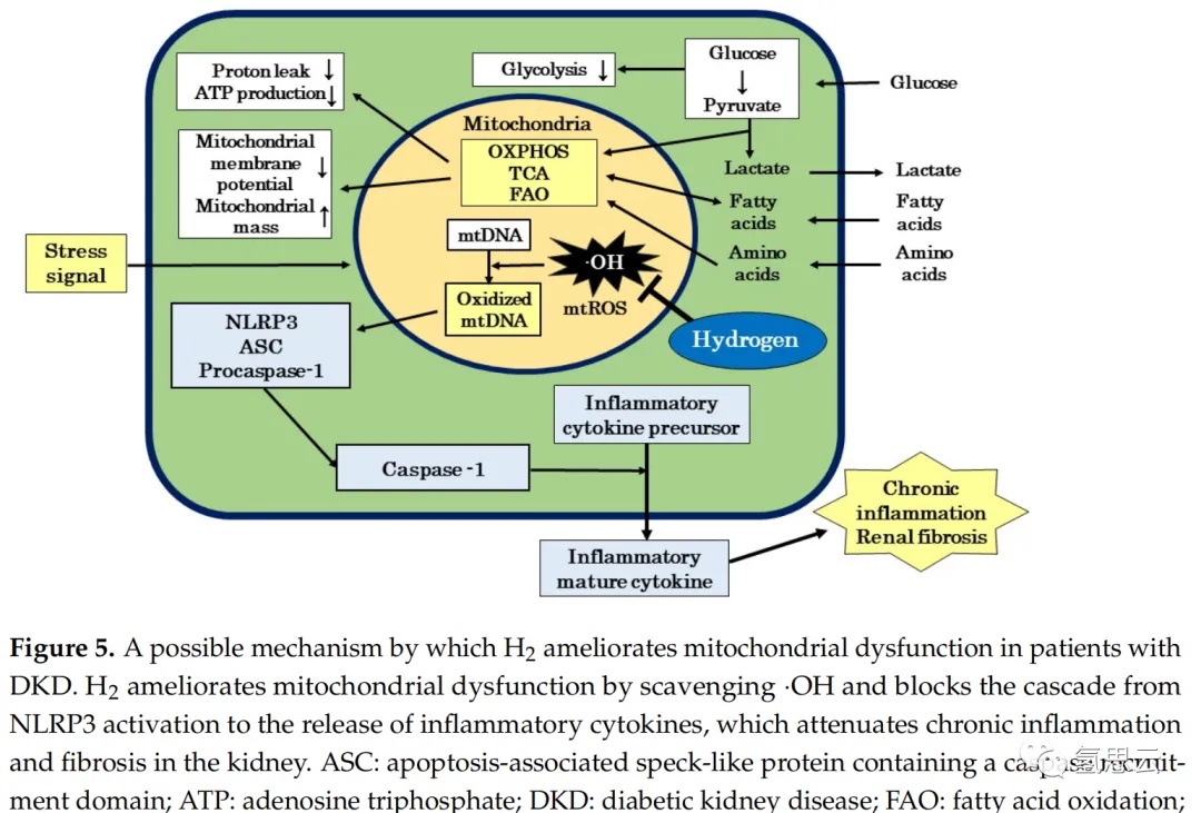
Inflammation is both a cause and a result of the occurrence and development of DKD. Inflammation is triggered by inflammatory factors released by the innate immune system. Pathogens such as viruses and bacteria, substances produced when the body is damaged, and stimuli in the environment all act as inflammatory induction signals. These external signals lead to mitochondrial dysfunction and excessive production of reactive oxygen species. The excessive production of mt reactive oxygen species in mitochondria leads to the release of oxidized mtDNA into the cytoplasm, which in turn leads to the generation of nucleotide binding and oligomerization domain like receptor family PYD domains containing protein 3 (NLRP3) inflammasome. Then NLRP3 inflammasome activates caspase-1, inducing immune cells (such as macrophages and neutrophils) to release mature inflammatory factors, leading to inflammation (Figure 5).
On the other hand, other intracellular oxidative stress responses are also involved in the induction of inflammation. The stress activated protein kinase (SAPK) pathway plays a core role in this oxidative stress response [124]. The SAPK pathway activated by oxidative stress stimuli induces the expression of various genes involved in stress response, ultimately leading to inflammation and cell death. In addition, reactive oxygen species play an important role in vascular inflammation. Excessive reactive oxygen species activate redox transcription factors such as nuclear factor kappa B (NF - κ B) and activating protein-1, leading to monocyte invasion of vascular walls and increased production of inflammatory cytokines. However, the presence of angiotensin II, oxidative LDL, and inflammatory cytokines activates NADPH oxidase, which leads to inflammation under oxidative stress conditions caused by excessive reactive oxygen species. Therefore, oxidative stress, inflammation, and endothelial dysfunction may be interrelated.
One of the risk factors for the progression of diabetes nephropathy (DKD) is the formation of interstitial damage. In renal tubulointerstitial injury associated with advanced proteinuria, excessive reabsorption of free fatty acids leads to renal tubular injury, which involves activating NLRP3 inflammasome through mitochondrial damage. In addition, the activation of mineralocorticoid receptors (MR) is closely related to renal inflammation and fibrosis, and it has been proven that MR activation induces the production of reactive oxygen species in MT. In addition, the activation of caspase-1 in glomerular epithelial cells may be important for the formation of glomerulosclerotic lesions in DKD. On the other hand, many studies have investigated the efficacy of hydrogen in inflammatory disease models, indicating that the mechanism by which hydrogen inhibits the production of reactive oxygen species in MT is involved in the inhibition of acute and chronic inflammation by hydrogen. Therefore, we propose a possible mechanism by which hydrogen inhibits the activation of NLRP3 inflammasome to release inflammatory cytokines through a series of effective signaling pathways by reducing ∙ OH and inhibiting oxidative damage to mtDNA. Hydrogen may reduce the formation of tubulointerstitial lesions in diabetes patients by inhibiting the activation of NLRP3 inflammatory body and improving chronic inflammation and renal fibrosis (Figure 5).
Diabetes peripheral neuropathy (DNP) is another serious complication of diabetes similar to DKD. Jiao et al. studied the effect of HRS on DNP in the model of streptozotocin induced diabetes rats, and found that it significantly inhibited the behavioral, biochemical and molecular biological effects of diabetes rats [136]. They also reported that the drug 5-hydroxydecanoic acid, which selectively inhibits mitochondrial ATP sensitive potassium channels (mitoKATP), can partially weaken the therapeutic effect of hydrogen saline. These findings indicate that the mechanism of action of hydrogen saline on DNP efficacy involves the protective effect on mitochondria by activating the mitoKATP pathway. In addition, we also reported that the effective mechanisms of hydrogen in certain animal disease models and human chronic inflammatory diseases (such as post COVID-19 sequelae known as post COVID-19 and myalgic encephalomyelitis/chronic fatigue syndrome (ME/CFS) may involve improvements in mitochondrial function. These findings suggest that hydrogen may be involved in improving mitochondrial function in the treatment of renal tubulointerstitial lesions in patients with diabetes (Figure 5).
7.2 Prospect of hydrogen as a therapeutic substance for diabetes nephropathy
Hydrogen has shown therapeutic effects in a wide range of diseases, and its efficacy has been reported in over 130 clinical papers. In these studies, no side effects of hydrogen were observed, therefore hydrogen is a medical gas with excellent efficacy and safety. In addition, hydrogen is a convenient gaseous molecule that can be directly inhaled as a gas, or dissolved in water or saline solution for drinking or intravenous injection. Hydrogen also exhibits excellent pharmacokinetic and intracellular kinetic properties. Mitochondrial dysfunction, oxidative stress, inflammation, and cell lethality are closely related to the onset and progression of DKD; Therefore, hydrogen may be effective for DKD. The mechanisms by which hydrogen has shown therapeutic effects in various animal kidney disease models and human dialysis patients, as well as our previous findings, provide evidence for the potential of hydrogen therapy for DKD. In addition, a literature review reviewing the effects of hydrogen on vascular endothelial function suggests that hydrogen may inhibit the progression of DKD to cardiovascular disease by inhibiting oxidative stress, inflammation, and apoptosis. Large scale clinical trials are still needed to demonstrate this potential.
On the other hand, research on the medical application of hydrogen has recognized some limitations. A recent study reported that porphyrin oxide serves as a target molecule for hydrogen and catalyzes the reaction between hydrogen and · OH [11]. However, the target effect molecules of hydrogen are still in the early stages and are only partially elucidated [11]. In addition, information on the dosage and use of individual diseases, including the optimal hydrogen concentration, daily dosage, and duration of intake, is still unclear. In addition, although hydrogen may have therapeutic effects on DKD by improving mitochondrial function, hydrogen may also involve other mechanisms. In addition, most DKD models used for animal research exhibit mild clinical symptoms, which are different from the clinical symptoms of human DKD. Therefore, it is necessary to further study the optimal dosage and usage of hydrogen for individual diseases, the mechanism of action of hydrogen, including its target molecules, and the development of animal models for DKD.
8. Conclusion
Hydrogen has shown therapeutic effects in various animal kidney disease models and dialysis patients. The mechanisms of action of hydrogen include mitochondrial improvement, antioxidant and anti-inflammatory effects, as well as regulation of cell lethality and intracellular signaling. Mitochondrial dysfunction, oxidative stress, inflammation, cell lethality, and intracellular signaling are all involved in the pathogenesis and progression of DKD. Our literature analysis of the efficacy of hydrogen gas in animal kidney disease models and human dialysis patients reported in this article suggests that hydrogen gas may have therapeutic potential in DKD patients. This therapeutic potential is supported by our mechanism analysis in this article, including the efficacy of hydrogen in human chronic inflammatory diseases such as COVID-19 sequelae and ME/CFS. Therefore, this review will provide an opportunity to consider the possibility of conducting clinical trials of hydrogen against DKD. Large scale clinical trials are needed in the future to confirm the impact of hydrogen on DKD.